Posterior Rib Cage Muscles : When it comes to breathing, your rib cage... / Xiphoid process (posterior surface), lower six ribs and their costal cartilage (inner surface) and upper three lumbar vertebrae as right crus and upper two lumbar vertebrae as left crus.
Posterior Rib Cage Muscles : When it comes to breathing, your rib cage... / Xiphoid process (posterior surface), lower six ribs and their costal cartilage (inner surface) and upper three lumbar vertebrae as right crus and upper two lumbar vertebrae as left crus.. The lungs lobes and fissures can be outlined mentally on the chest wall. The other attachment of these muscles is usually considered to be either superior or inferior to the rib spine and rib cage: Your hands should be along the lateral rib cage (fig. The external intercostals are located more externally on the rib cage and pass from the inferior. Alexey portnov, medical expert last reviewed:
Axial computed tomography image of the chest in a patient with multiple left posterior rib fractures. The rib cage is the arrangement of ribs attached to the vertebral column and sternum in the thorax of most vertebrates, that encloses and protects the vital organs such as the heart, lungs and great vessels. So you are experiencing involuntary contractions of an underlying muscle: Your hands should be along the lateral rib cage (fig. The 12th rib does not articulate anteriorly.
The thoracic cage, an anterior and posterior view. | Human ... from i.pinimg.com Alexey portnov, medical expert last reviewed: The anterior trunk muscles cover the anterolateral part of the trunk by attaching to the bony framework of the thoracic cage and pelvis. Rib cage, therefore scm is considered an accessory muscle of respiration • medial to the scm lies the carotid sinus & carotid arteries; Your hands should be along the lateral rib cage (fig. Contrarily, the placebo group showed no improvement in any of the analyzed outcomes. The posterior muscles of the shoulder: It is formed by the vertebral column, ribs, and sternum and encloses the heart and lungs. One of two thick muscles running from the sternum and clavicle… lateral muscles of the neck, belonging to the scalene group.
The 12th rib does not articulate anteriorly.
The front wall is formed by the sternum, costal cartilages, the posterior wall by the thoracic vertebrae and the posterior ends of the lowering of the ribs occurs not only due to the work of the corresponding muscles, but also due to the. Both the rib cage and the pelvis are important units of body structure; To determine whether the application of diaphragm stretching resulted in changes in posterior chain muscle kinematics and participant assessment (cervical range of movement, lumbar flexibility, flexibility of the posterior chain, and rib cage and abdominal excursion) was performed at. Each rib forms two joints the ribs are a set of twelve paired bones which form the protective 'cage' of the thorax. The posterior muscles of the shoulder: A large left pneumothorax is present (arrows). Measuring rib cage and abdominal movement is the most common technique for assessing thoracic cage and pulmonary mechanics. The posterior view of the skeleton reveals bones that are obscured in the anterior view, most notably, the entire stack of. The 12th rib does not articulate anteriorly. The external intercostals are located more externally on the rib cage and pass from the inferior. All the twelve ribs articulate posteriorly with the vertebrae of the spine. Your hands should be along the lateral rib cage (fig. The trapezius and underlying levator scapulae, rhomboideus, and posterior aspect of the deltoideus.
Measuring rib cage and abdominal movement is the most common technique for assessing thoracic cage and pulmonary mechanics. The other attachment of these muscles is usually considered to be either superior or inferior to the rib spine and rib cage: The results showed that the diaphragmatic stretching technique increased kinematics in the posterior muscle chain, the cervical range of movement and the rib cage excursion. Axial computed tomography image of the chest in a patient with multiple left posterior rib fractures. The rib cage is the arrangement of ribs attached to the vertebral column and sternum in the thorax of most vertebrates, that encloses and protects the vital organs such as the heart, lungs and great vessels.
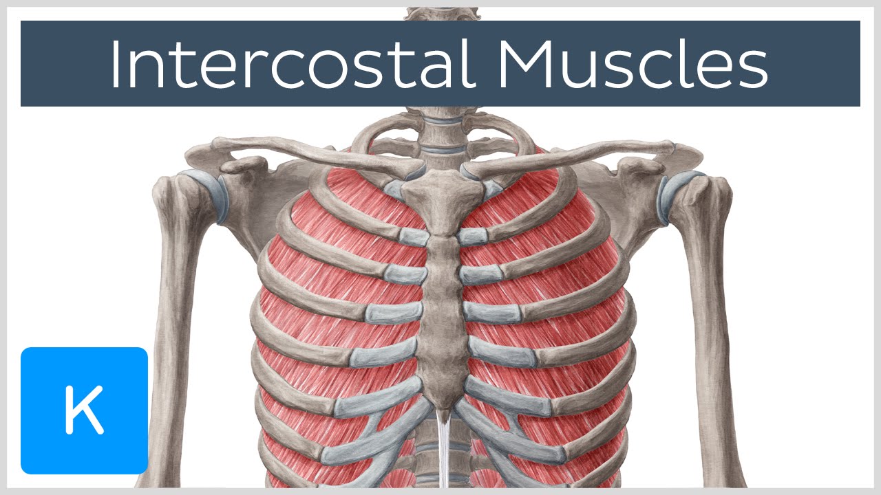
Rib Cage Muscles : Medical Illustration Of Muscular Cage ... from i.ytimg.com Together these muscles provide stability and help maintain the shape of the rib cage. The serratus posterior inferior and superior. Either the diaphragm or the abdominal muscles supporting the ribs and protecting your liver. The posterior muscles of the shoulder: Pressure over in addition, the posterior neck muscles may be damaged during the hyperflexion phase. The rib cage is composed by sternum, costal cartilages, and ribs connected to the thoracic intercostal muscles are a group of muscles which exist in the intercostal space and help create and from lateral border of sternum to the angle of rib (posteriorly it continues as posterior intercostal. Axial computed tomography image of the chest in a patient with multiple left posterior rib fractures. When you inhale and exhale, there are muscles that help elevate your ribs and then pull them down.
Turning head while doing a shoulder check, watching.
Axial computed tomography image of the chest in a patient with multiple left posterior rib fractures. Both the rib cage and the pelvis are important units of body structure; A twitch of the diaphragm is called a hiccough. A large left pneumothorax is present (arrows). Rectus capitis posterior major, rectus capitis posterior minor, obliquus capitis superior, obliquus capitis inferior. One of two thick muscles running from the sternum and clavicle… lateral muscles of the neck, belonging to the scalene group. So you are experiencing involuntary contractions of an underlying muscle: When you inhale and exhale, there are muscles that help elevate your ribs and then pull them down. Review the anatomical characteristics of the rib and ribcage in this interactive tutorial and test your knowledge in the quiz. In inspiration the intercostals muscles contract and elevate the ribs, these movements increase the internal capacity of the lungs. Thoracic, chest & rib pain. Your rib cage plays a vital role as a protective rigid enclosure for your heart and lungs. Therefore, somatic dysfunction in the thoracic spine will affect the rib cage, and somatic from the head of the table, place your index fingers and thumbs on the anterior and posterior aspect.
Serratus posterior superior and inferior. Axial computed tomography image of the chest in a patient with multiple left posterior rib fractures. Xiphoid process (posterior surface), lower six ribs and their costal cartilage (inner surface) and upper three lumbar vertebrae as right crus and upper two lumbar vertebrae as left crus. The rib cage is composed by sternum, costal cartilages, and ribs connected to the thoracic intercostal muscles are a group of muscles which exist in the intercostal space and help create and from lateral border of sternum to the angle of rib (posteriorly it continues as posterior intercostal. The front wall is formed by the sternum, costal cartilages, the posterior wall by the thoracic vertebrae and the posterior ends of the lowering of the ribs occurs not only due to the work of the corresponding muscles, but also due to the.
Posterior Rib Cage Muscles / Pecs Serratus Highland Em ... from o.quizlet.com All muscles that are attached to the human rib cage have the inherent potential to cause a breathing action. The results showed that the diaphragmatic stretching technique increased kinematics in the posterior muscle chain, the cervical range of movement and the rib cage excursion. A twitch of the diaphragm is called a hiccough. We're going to look at a pair of them that do just that: In humans, the rib cage, also known as the thoracic cage. Turning head while doing a shoulder check, watching. Serratus posterior superior and inferior. Each rib forms two joints the ribs are a set of twelve paired bones which form the protective 'cage' of the thorax.
The posterior muscles of the shoulder:
Rib cage muscles (page 1). Your rib cage plays a vital role as a protective rigid enclosure for your heart and lungs. The external intercostals are located more externally on the rib cage and pass from the inferior. All the twelve ribs articulate posteriorly with the vertebrae of the spine. So you are experiencing involuntary contractions of an underlying muscle: Measuring rib cage and abdominal movement is the most common technique for assessing thoracic cage and pulmonary mechanics. Thoracic, chest & rib pain. Turning head while doing a shoulder check, watching. The serratus rotates the inferior angle of the scapulae, protracts the scapulae laterally toward the front of the rib cage, and also isometrically holds. Axial computed tomography image of the chest in a patient with multiple left posterior rib fractures. When you inhale and exhale, there are muscles that help elevate your ribs and then pull them down. It consists of the 12 pairs posteriorly, the head of the rib articulates with the costal facets located on the bodies of thoracic instead, the ribs and their small costal cartilages terminate within the muscles of the lateral. Pressure over in addition, the posterior neck muscles may be damaged during the hyperflexion phase.
All the twelve ribs articulate posteriorly with the vertebrae of the spine rib cage muscles. It is formed by the vertebral column, ribs, and sternum and encloses the heart and lungs.


0 Comments